The normal microscopic appearance of the pituitary gland is shown above. The pituitary is composed of the.

Endocrine Systems Lab
It connects to the pituitary gland by the stalk-like infundibulum.

. The anterior lobe appears darker. Examine a section of pituitary slide 128 103 and identify the anterior pituitary the posterior pituitary and the intervening pars intermedia with cysts representing remnants of Rathkes pouch Figs. Anterior and posterior pituitary hormones.
Anterior Gastrocnemius Soleus Tibialis Fibularis Longus Extensor Digitorum Longus Tendon of Gastrocnemius. This gland is located just below the brain somewhat behind the eyes. Make a sketch of your observed field of view and label.
As the human embryo develops the anterior pituitary is formed from cells from the roof of the mouth that migrate toward the brain. The pituitary gland is called the master gland as the hormones it produces control so many different processes in the body. Draw in the location of the hypothalamus and pineal gland with respect to pituitary gland.
The anterior and posterior pituitary glands are the two components of the pituitary gland. Identify the pineal gland on a histology slide. The pituitary gland is split into two different portions and the anterior is at the front while the posterior is in the back.
Hope this guide was helpful to learn pituitary gland histology. Anterior pituitary produces its own hormones while posterior pituitary stores hormones which are originally produced in the hypothalamus. Slide of Pituitary Gland In the space below draw and label the anterior pituitary gland and the posterior pituitary gland.
Precise spatial and temporal co-ordination of transcription factor expression in both structures is critical for pituitary formation and the differentiation of. Pituitary Gland anterior - 400X. The anterior lobe adenohypophysis stains darker and the posterior lobe neurohypophysis stains lighter.
A What hormones from the hypothalamus will. The posterior part of the pituitary has its embryological origins in nervous tissue. The posterior pituitary pars nervosa is connected to the hypothalamus by the pituitary stalk - this is easily visualized because the nervous tissue appears continuous between the two glands.
Terminologies for the components of the pituitary gland are based on the embryological origins of the main subdivisions as well as the anatomical regions of each. What is the difference between Anterior and Posterior Pituitary. Describe the structure and functions of pinealoytes and pineal sand.
The anterior pituitary receives signaling molecules from the hypothalamus and in response synthesizes and secretes seven important hormones including thyroid-stimulating hormone and growth hormone. In the anterior pituitary pars distalis you can see cords of cuboidal cells with a wide range of nuclear to cytoplasmic volume ratios. Each of these parts of the pituitary gland do very different but equally important things.
The pituitary gland is split into two different portions and the anterior is at the front while the posterior is in the back. Anterior pituitary disorder characterized by. Now you will able to identify the anterior and posterior pituitary gland histology slide under microscope with important identification points.
The production and release of hormones by the anterior and posterior pituitary glands are regulated by the hormones. The hypothalamus regulates the pituitary gland secretion12343 The pituitary endocrine gland which is located in the bony sella. The pituitary gland is actually composed of two glands.
Anterior pituitary is derived from ectodermal tissue whereas posterior pituitary is derived from the ventral surface of the diencephalon. The pituitary gland consists of an anterior and posterior lobe with each lobe secreting different hormones. PITUITARY GLAND Has two parts.
LEG MUSCLE MODEL Tendon of Gluteus. Both anterior and posterior pituitary glands are endocrine glands releasing hormones to regulate the functions of most of the organs and glands in the body. The pars intermedia is poorly developed in humans.
Anterior pituitary glandular Posterior pituitary neural Alternative names. The hypothalamus region lies inferior and anterior to the thalamus. Obtain a prepared slide of the pituitary gland Begin observing it under scanning power and notice the difference between the anterior pituitary and posterior pituitary tissue organization.
The anterior part is derived from an upgrowth from the oral ectoderm of the primitive oral cavity called Rathkes pouch. The pituitary is an organ dual of origin. Between these lobes lies a small region called the intermediate lobe.
The pituitary gland sometimes called the hypophysis is a small gland that dangles from the base of the brain like a pea on a string Several hormones produced by the hypothalamus are stored here and released into the blood. The posterior lobe secretes hormones that are made by the hypothalamus. Switch to low power and observe the area that separates the two parts as shown in Figure 113.
In clinical reference the term anterior pituitary is often used synonymously with pars distalis and posterior pituitary is frequently used synonymously with pars nervosa. When the structure and function of the pituitary glands are impacted by tumors trauma or genetic disorders it can cause a variety of medical conditions including hypopituitarism prolactinoma and Cushings disease. Differentiate between the anterior and posterior lobes of the pituitary gland on a histology slide.
The posterior pituitary is composed of neural tissue. Through secretion of its hormones the pituitary gland controls metabolism growth sexual maturation reproduction blood pressure and many other vital physical functions and processes. The anterior and posterior pituitary glands are the two components of the pituitary gland.
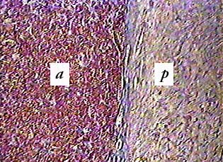
A anterior lobe b posterior lobe. If you look closely you can see that the cells are arranged in clusters. Dont forget to join social media of anatomy learner for more update pituitary gland histology labeled pictures and diagram.
The pituitary gland consists of two anatomically and functionally distinct regions the anterior lobe adenohypophysis and the posterior lobe neurohypophysis. It is formed from a downgrowth of the diencephalon that forms the floor of the third ventricle. Adenohypophysis pars distalis pars anterior.
This is the view under a dissecting microscope. Abnormal growth of the hands feet and face caused by overproduction of growth hormone by the pituitary gland. This slide shows a section of the human pituitary.
If you cant see the anterior and posterior pituitary glands next to each other on a slide they both become much harder to identify. Enlargement thickening and broadening of bones Particularly extremities of the body. Pars intermedia separates anterior from posterior.
The pituitary gland is divided into two parts the anterior pituitary and the posterior pituitary. Along the posterior part of the the anterior lobe there is a narrow region called. The anterior pituitary can be confused with the pancreas or parotid glands or even the liver.
The anterior pituitary the posterior pituitary. The latter structure may be better seen on slide 122. PITUITARY GLAND SLIDE Posterior Lobe Anterior Lobe _____ _____ _____ _____ _____ THYROID GLAND SLIDE Parafollicular Cells Colloid fluid within the follicle.
The anterior lobe adenohypophysis is epithelial in origin whereas the posterior lobe neurohypophysis derives from the neural ectoderm. The anterior and posterior pituitary glands release different sets of hormones.
Pin On Histology

Pin On Histology A P Ii

Pin On Histology A P Ii

Endocrine Systems Lab

Endocrine Systems Lab
Pituitary Gland

A P Ii Endocrine Gland Slides Flashcards Quizlet

Pituitary Left Anterior Right Posterior Ppt Download
0 comments
Post a Comment